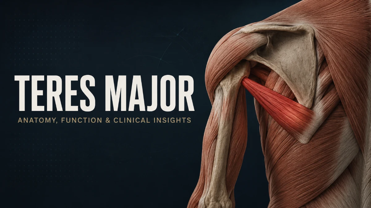
The teres major is a posterior shoulder muscle that runs from the lower part of the scapula to the upper humerus. It helps extend, adduct, and internally rotate the arm, working closely with the latissimus dorsi during pulling, rowing, climbing, swimming, and throwing movements. Although it lies near the teres minor, it is not part of the rotator cuff.
Understanding this muscle is useful for anatomy students, fitness professionals, healthcare learners, and anyone researching pain behind the shoulder or near the back of the armpit. This guide explains the teres major muscle anatomy, origin and insertion, actions, related pain patterns, injury considerations, and clinical importance.
Teres Major Quick Anatomy Summary
| Feature | Detail |
| Muscle group | Scapulohumeral muscle |
| Location | Posterior shoulder and posterior wall of the axilla |
| Origin | Posterior surface of the inferior angle of the scapula and adjacent lower lateral border |
| Insertion | Medial lip of the intertubercular sulcus of the humerus, also called the crest of the lesser tubercle |
| Main actions | Shoulder extension, adduction, and internal rotation |
| Primary nerve | Lower subscapular nerve from the posterior cord of the brachial plexus |
| Main functional partner | Latissimus dorsi |
| Rotator cuff muscle | No |
| Important regional relationship | Forms part of the inferior border of the quadrangular space |
What Is the Teres Major Muscle?
The teres major is a thick, flattened muscle on the back of the shoulder. It begins on the lower part of the shoulder blade and travels upward and laterally to the humerus. When it contracts, it pulls the upper arm backward, toward the trunk, and inward.
It is classified as a scapulohumeral muscle because it connects the scapula to the humerus and acts directly at the glenohumeral joint. The muscle lies inferior to the teres minor and contributes to the posterior wall of the axilla. Its position is important in posterior shoulder anatomy because it is close to the long head of the triceps brachii, latissimus dorsi, axillary nerve, and posterior circumflex humeral artery.
Read more: Teres Major
Where Is the Teres Major Located?
The muscle occupies the lower back portion of the shoulder. It begins near the inferior angle of the scapula, passes toward the upper arm, and helps form the posterior axillary fold with the latissimus dorsi.
Its anatomical relationships include:
- Superiorly: Teres minor
- Inferiorly: Latissimus dorsi and posterior axillary fold
- Medially: Inferior angle and lateral border of the scapula
- Laterally: Proximal humerus near the intertubercular groove
- Nearby: Long head of the triceps brachii and posterior shoulder structures
In people with developed back musculature, it may be visible or palpable as part of the muscular contour behind the armpit during resisted shoulder extension or adduction.
Teres Major Origin and Insertion
The teres major originates from the posterior surface of the inferior angle of the scapula and the adjacent lower part of the scapula’s lateral border. Its fibres travel upward and laterally, then converge into a short tendon.
The tendon inserts onto the medial lip of the intertubercular sulcus of the humerus, also known as the crest of the lesser tubercle. This insertion lies close to the attachment areas of the latissimus dorsi and pectoralis major.
| Attachment | Anatomical detail | Functional importance |
| Origin | Posterior surface of the inferior angle and lower lateral border of the scapula | Establishes a pull from scapula to humerus |
| Insertion | Medial lip of the intertubercular groove of the humerus | Allows movement at the glenohumeral joint |
| Fibre direction | Upward and lateral | Supports extension, adduction, and internal rotation |
| Tendon relationship | Close to latissimus dorsi and pectoralis major insertions | Explains overlapping pulling actions |
Because the tendon attaches on the anterior aspect of the proximal humerus after crossing from the posterior scapula, its contraction contributes to internal rotation. Its lower and medial line of pull also supports adduction and extension.
Teres Major Function
The primary functions of the teres major are shoulder extension, adduction, and internal rotation. It works most noticeably when the arm pulls downward, backward, or toward the body.
Main Actions of the Teres Major
- Shoulder extension: Moves the arm backward from a flexed or overhead position.
- Shoulder adduction: Draws the upper arm toward the side of the trunk.
- Internal rotation: Rotates the humerus toward the body’s midline.
- Dynamic movement control: Assists coordinated movement at the glenohumeral joint during pulling and overhead activity.
The teres major is not a rotator cuff muscle and is not generally considered a primary humeral-head stabilizer in the same way as the supraspinatus, infraspinatus, teres minor, and subscapularis. However, it contributes to efficient shoulder movement by working with the latissimus dorsi, subscapularis, deltoid, scapular stabilizers, and trunk muscles.
Everyday and Athletic Movements
This muscle contributes to:
- Pull-ups and chin-ups
- Lat pulldowns
- Straight-arm pulldowns
- Cable and dumbbell rows
- Swimming pull phases
- Rock climbing
- Throwing follow-through
- Reaching down from an elevated position
- Pulling an object toward the body
The teres major may be exposed to high eccentric load during forceful overhead throwing, when the arm must be controlled after ball release. It can also be stressed during sudden, forceful pulling tasks.
Nerve and Blood Supply
The teres major is usually supplied by the lower subscapular nerve, a branch of the posterior cord of the brachial plexus. The nerve typically carries fibres from the C5 and C6 spinal nerve roots.
The lower subscapular nerve also innervates part of the subscapularis. This relationship matters clinically because injury affecting the posterior cord or lower subscapular nerve can impair more than one shoulder internal-rotation muscle.
Anatomical variations in nerve supply have been reported, including possible contributions from the thoracodorsal nerve. These patterns are not universal but may be relevant in detailed anatomical dissection and surgical planning.
Blood Supply
The muscle receives arterial blood through branches of the axillary artery system, particularly branches related to the subscapular artery. The thoracodorsal and circumflex scapular arterial systems are important regional contributors.
Teres Major and Shoulder Anatomy
The teres major acts directly at the glenohumeral joint, but its performance depends on the entire shoulder complex. This includes the scapulothoracic articulation, clavicle, thorax, rotator cuff, deltoid, and scapular stabilizers.
The scapulothoracic articulation is the functional relationship between the scapula and the thoracic wall rather than a true synovial joint. Controlled scapular upward rotation, posterior tilt, and positioning help create a stable base for arm movement.
Important Anatomical Relationships
| Structure | Relationship to the teres major |
| Latissimus dorsi | Shares extension, adduction, and internal-rotation actions |
| Teres minor | Lies superiorly; externally rotates the arm and belongs to the rotator cuff |
| Long head of triceps brachii | Runs near the muscle in the posterior shoulder region |
| Subscapularis | Shares internal-rotation function and lower subscapular nerve supply |
| Deltoid | Covers portions of the posterior shoulder region superficially |
| Axillary nerve | Passes through the quadrangular space near the upper border region of teres major |
| Posterior circumflex humeral artery | Travels with the axillary nerve through the quadrangular space |
| Scapular stabilizers | Position the scapula and influence shoulder mechanics |
Quadrangular Space Relationship
The teres major forms the inferior border of the quadrangular space. The teres minor forms the superior border, the long head of the triceps brachii forms the medial border, and the surgical neck of the humerus forms the lateral border.
The axillary nerve and posterior circumflex humeral artery pass through this space. This relationship is important when assessing posterior shoulder trauma, nerve compression, or anatomy during surgical procedures.
Teres Major vs. Teres Minor
The teres major and teres minor have similar names and lie near each other, but they have different attachments, actions, and innervation.
| Feature | Teres Major | Teres Minor |
| Location | Lower posterior shoulder | Upper posterior shoulder |
| Origin | Inferior angle and lower lateral border of scapula | Lateral border of scapula |
| Insertion | Medial lip of intertubercular groove | Greater tubercle of humerus |
| Main action | Extension, adduction, internal rotation | External rotation |
| Nerve supply | Lower subscapular nerve | Axillary nerve |
| Rotator cuff member | No | Yes |
| Typical clinical relevance | Pulling-related pain and posterior axillary symptoms | Rotator cuff dysfunction and external-rotation weakness |
The clearest functional difference is rotation: the teres major internally rotates the arm, while the teres minor externally rotates it. The teres minor is also one of the four rotator cuff muscles, while the teres major is not.
Teres Major vs. Latissimus Dorsi
The teres major and latissimus dorsi have overlapping functions and are frequently activated together during pulling movements. However, the latissimus dorsi is a much larger muscle with broader connections to the trunk and pelvis.
| Feature | Teres Major | Latissimus Dorsi |
| Size | Smaller posterior shoulder muscle | Large superficial back muscle |
| Main origin | Inferior angle and lower lateral border of scapula | Thoracolumbar fascia, lower thoracic vertebrae, lumbar spine, iliac crest, and lower ribs |
| Insertion | Medial lip of intertubercular groove | Floor of the intertubercular groove |
| Main actions | Extension, adduction, internal rotation | Extension, adduction, internal rotation |
| Trunk involvement | Limited | Transfers force between upper limb and trunk |
| Injury pattern | Less commonly reported | More often described in high-demand throwing athletes |
| Common pain area | Posterior axilla and lower shoulder | Side of back and posterior axilla |
Because both muscles can produce posterior axillary pain and participate in forceful pulling, it can be difficult to identify the involved structure without a clinical assessment. The two muscles are also often considered together during imaging evaluation because of their close anatomical relationship.
Teres Major Pain and Injuries
Teres major injuries are less common than rotator cuff disorders, biceps tendon conditions, or broader latissimus dorsi injuries. When they occur, they may involve a mild strain, a myotendinous injury, a partial tear, or, rarely, a complete rupture.
They are most often associated with high-force movements that involve shoulder extension, adduction, and internal rotation. Throwing, climbing, water skiing, rowing, swimming, bodybuilding, and heavy pulling may create this type of loading.
Common Causes of Teres Major Pain
- Sudden forceful pulling movement
- Repetitive overhead throwing
- Rapid arm deceleration after ball release
- Heavy pull-ups, rows, or pulldowns
- Direct trauma to the posterior shoulder or axillary region
- Fatigue-related movement changes during sport or resistance training
Where Is Teres Major Pain Felt?
Pain related to this muscle is often felt in the lower posterior shoulder, the posterior axilla, or along the lower outer border of the scapula. Some people describe it as discomfort under the shoulder blade or behind the armpit.
Possible symptoms include:
- Local tenderness near the posterior axillary fold
- Pain with resisted shoulder extension or adduction
- Discomfort during pulling, climbing, rowing, or throwing
- Weakness during forceful shoulder movements
- Pain when lowering the arm from an overhead position
- Bruising or swelling after a substantial acute injury
Pain location alone cannot confirm a teres major strain. Similar symptoms can arise from the latissimus dorsi, rotator cuff, long head of triceps, cervical spine, scapular muscles, or nerve-related conditions.
Teres Major Pain vs. Similar Shoulder Conditions
Posterior shoulder pain may involve more than one structure. The table below outlines common symptom patterns, but it cannot replace a clinical diagnosis.
| Condition | Typical pain location | Movements that may trigger pain | Distinguishing features |
| Teres major strain | Posterior axilla, lower posterior shoulder, lower scapular border | Pulling, shoulder extension, adduction, internal rotation | Often follows a forceful pull or throwing event |
| Latissimus dorsi strain | Side of the back, posterior axilla, lower shoulder | Powerful pulling, throwing, swimming | Pain may extend more broadly into the side or back |
| Rotator cuff disorder | Lateral or front-lateral shoulder | Overhead reaching, lifting, sleeping on the shoulder | Painful arc, weakness, and overhead symptoms may be more prominent |
| Teres minor injury | Posterior shoulder | Resisted external rotation | External-rotation weakness may be present |
| Long-head triceps pathology | Posterior upper arm or shoulder | Resisted elbow extension or overhead activity | Tenderness may follow the triceps course |
| Cervical radiculopathy | Neck, shoulder, arm, or hand | Neck movement can aggravate symptoms | Tingling, numbness, radiating pain, or reflex changes may occur |
A clinician may use history, palpation, range-of-motion testing, resisted movement testing, strength comparison, and a neurological examination to narrow the diagnosis. Imaging may be considered when symptoms are severe, persistent, associated with bruising or weakness, or suggest a substantial tear.
If a teres major or latissimus dorsi injury is suspected, the imaging request should clearly state that concern. A standard shoulder MRI may not include the full posterior axillary region or the entire muscle-tendon pathway.
Teres Major Strain Recovery and Rehabilitation
Recovery depends on injury severity, tissue involved, physical demands, and individual health factors. A return to work, training, or sport should be based on functional recovery rather than a fixed timeline.
| Injury pattern | General description | Rehabilitation focus |
| Mild strain | Small amount of muscle fibre damage with pain but minimal strength loss | Symptom control, early mobility, gradual strengthening |
| Moderate strain or partial tear | Greater tissue disruption, pain, weakness, bruising, or reduced function | Structured rehabilitation and possible imaging |
| Complete tear | Full disruption of the muscle or tendon, sometimes with retraction | Specialist assessment and individualized management |
Early Symptom-Control Phase
The early focus is reducing pain while avoiding movements that repeatedly stress the injured tissue.
- Use relative rest and avoid movements that cause sharp or worsening pain.
- Pause throwing, pull-ups, heavy rows, pulldowns, climbing, and forceful overhead pulling.
- Maintain comfortable movement of the elbow, wrist, neck, and pain-free shoulder ranges.
- Seek assessment if weakness, bruising, deformity, or major loss of function occurs.
Mobility Restoration Phase
Once pain begins to settle, the aim is to restore comfortable shoulder and scapular movement.
- Gradually restore pain-free shoulder flexion, extension, and rotation.
- Improve scapular control and thoracic mobility where appropriate.
- Avoid aggressive stretching that reproduces sharp posterior shoulder or axillary pain.
- Progress based on movement quality and symptom response.
Progressive Strengthening Phase
Strengthening should begin with low-load, controlled exercise and advance only when it is well tolerated.
- Begin with appropriate isometric shoulder extension and adduction exercises.
- Progress to light rows, resistance-band pulldowns, and controlled internal-rotation exercises.
- Include scapular stabilizer training to support efficient shoulder mechanics.
- Increase load gradually and monitor pain during and after exercise.
Return-to-Training Phase
A return to sport or demanding training should occur when the person can complete relevant movements without pain, weakness, or compensation.
Key return-to-training markers include:
- Full or near-full pain-free shoulder range of motion
- No increasing tenderness after exercise
- Comparable strength and control between sides
- Tolerance of progressive pulling or overhead work
- Ability to complete sport-specific drills without symptoms
- Clinical clearance after a significant tear or in high-demand athletes
When to Seek Medical Care
Seek timely medical assessment for:
- Sudden severe pain after throwing, pulling, falling, or trauma
- A pop sensation followed by bruising, swelling, or visible contour change
- Significant weakness when pulling the arm downward or toward the body
- Persistent posterior shoulder pain that does not improve with activity modification
- Numbness, tingling, neck pain, or pain radiating into the arm
- Difficulty returning to work, exercise, or sport
- Suspected tendon rupture or substantial muscle tear
Clinical and Surgical Importance
The teres major is an important landmark in posterior shoulder and axillary anatomy because of its relationship with the quadrangular space, latissimus dorsi, long head of the triceps, lower subscapular nerve, and nearby vessels.
In selected reconstructive cases, the muscle has been studied as a tendon-transfer option for irreparable subscapularis tears. This is a specialised approach rather than a standard treatment for routine shoulder pain or ordinary muscle strain. Surgical decisions depend on the specific tear pattern, tendon quality, nerve function, shoulder arthritis, prior procedures, functional needs, and overall health.
Teres Major Exercises
The muscle is usually trained as part of compound pulling patterns rather than with complete isolation. These exercises recruit it alongside the latissimus dorsi, deltoid, triceps, rotator cuff, and scapular stabilizers.
| Exercise | How it recruits the muscle |
| Exercise | How it recruits the muscle |
| Pull-ups | Assists shoulder adduction and extension |
| Lat pulldowns | Supports arm adduction during the pulling phase |
| Straight-arm pulldowns | Emphasizes shoulder extension |
| Cable rows | Assists humeral extension while the shoulder blade is stabilized |
| Dumbbell rows | Helps move the upper arm backward toward the torso |
| Swimming pull phase | Assists extension and adduction of the arm |
| Resistance-band internal rotation | Trains internal rotation, although not in isolation |
Use controlled technique and progressive loading. Avoid training through sharp pain, sudden weakness, or persistent symptoms near the posterior shoulder or axilla.
Frequently Asked Questions
Is the teres major part of the rotator cuff?
No. The rotator cuff consists of the supraspinatus, infraspinatus, teres minor, and subscapularis. The teres major lies close to the teres minor but is a separate scapulohumeral muscle. It helps extend, adduct, and internally rotate the arm, while the teres minor contributes mainly to external rotation. The teres major supports coordinated shoulder movement but is not classified as a rotator cuff muscle.
Where exactly is the teres major muscle located?
It lies in the lower posterior shoulder. The muscle begins at the inferior angle and lower lateral border of the scapula, then travels toward the proximal humerus. It contributes to the posterior axillary fold with the latissimus dorsi. People often notice this region as the muscular area behind the armpit or along the lower outer edge of the shoulder blade.
What causes teres major pain?
Pain may develop after heavy pulling, throwing, climbing, swimming, rowing, direct trauma, or rapid arm deceleration. A strain can affect the muscle belly, the myotendinous junction, or the tendon. However, pain in this region can also come from the latissimus dorsi, rotator cuff, long head of triceps, neck, or nerve irritation. Persistent pain, bruising, weakness, or loss of function warrants clinical assessment.
How can teres major pain be distinguished from rotator cuff pain?
Pain from the teres major is often lower and more posterior, near the back of the armpit or lower scapular border. It may worsen with resisted pulling, shoulder extension, adduction, or internal rotation. Rotator cuff symptoms more often occur at the side of the shoulder and may worsen with overhead reaching, lifting away from the body, or lying on the affected side. The patterns can overlap, so diagnosis requires a clinical examination.
How long does a teres major strain take to heal?
Healing time varies with the degree of muscle or tendon injury. A mild strain may improve over several weeks, while partial or complete tears can require a much longer and more structured recovery. Progress should be based on restored range of motion, strength, and tolerance of work or sport tasks rather than a fixed timeframe. A clinician should assess substantial weakness, bruising, or symptoms that do not improve.
Can a teres major tear be seen on MRI?
MRI can identify muscle edema, partial tears, complete tears, and tendon retraction. However, a routine shoulder MRI may not include the entire teres major because the muscle extends into the posterior axillary region. If this injury is suspected, the referring clinician should specify the concern so the imaging protocol can include an appropriate field of view and targeted image planes.
Which exercises should be avoided with a teres major strain?
Avoid movements that reproduce pain or heavily load shoulder extension, adduction, and internal rotation. This often includes pull-ups, lat pulldowns, heavy rows, straight-arm pulldowns, climbing, swimming pull phases, and throwing. Relative rest is usually preferable to complete inactivity. Resume these activities gradually only when movement is comfortable and strength has returned, ideally with guidance from a qualified healthcare or rehabilitation professional.
Does the teres major help with pull-ups?
Yes. It assists shoulder adduction and extension during pull-ups as the upper arm moves down toward the trunk. It works with the latissimus dorsi, biceps, lower trapezius, rhomboids, rotator cuff, and other shoulder muscles. Although it is not usually the sole limiting muscle in this exercise, it contributes to the pulling pattern and may become overloaded when training volume or load rises too quickly.
Can a tight teres major limit overhead movement?
It may contribute to discomfort or limited overhead movement when combined with restricted latissimus dorsi flexibility, reduced thoracic mobility, altered scapular mechanics, or other shoulder impairments. However, overhead stiffness and impingement-type symptoms are usually multifactorial. It is not accurate to attribute persistent shoulder restriction to the teres major alone without considering the entire shoulder and cervical spine.
Why is the teres major clinically important in shoulder surgery?
Its location makes it an important landmark near the axillary nerve, posterior circumflex humeral artery, lower subscapular nerve, and posterior shoulder muscles. It has also been studied for use in highly selected tendon-transfer procedures for irreparable subscapularis tears. These operations are specialised and are not treatments for routine muscle soreness or uncomplicated strains; an orthopedic shoulder surgeon determines whether they are appropriate.
