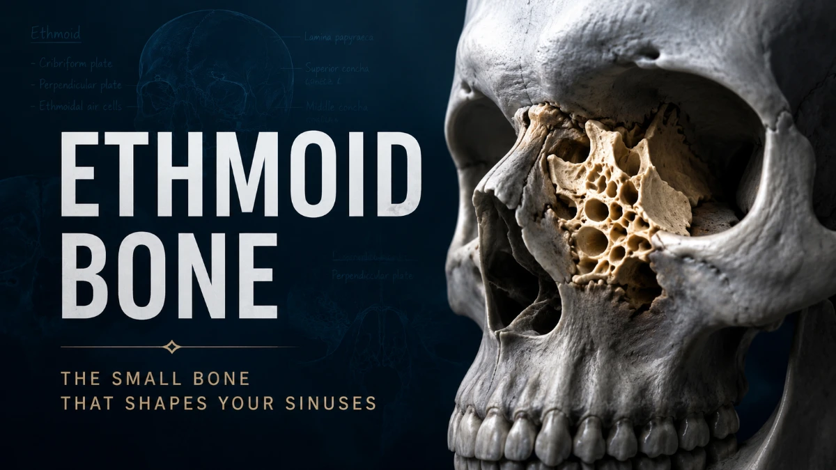
The ethmoid bone is a small, unpaired cranial bone located between the eyes and behind the bridge of the nose. It forms parts of the nasal cavity, medial orbital walls, anterior skull base, and ethmoid sinuses. Its cribriform plate also transmits smell-related nerve fibers from the nasal cavity to the olfactory bulbs.
Ethmoid Bone Quick Facts
| Feature | Detail |
| Classification | Unpaired cranial bone |
| Location | Between the orbits, above the nasal cavity, beneath the frontal bone |
| Main parts | Cribriform plate, crista galli, perpendicular plate, paired ethmoidal labyrinths |
| Major functions | Supports olfaction, contributes to nasal structure, contains ethmoid air cells, and helps form the orbit and skull base |
| Key nerve relationship | Olfactory nerve, cranial nerve I |
| Key orbital structure | Lamina papyracea |
| Clinical relevance | Sinus disease, facial trauma, skull-base anatomy, orbital complications, and endoscopic sinus surgery |
Why the Ethmoid Bone Is Unique
The ethmoid is unusually important for such a small bone because it forms a junction between the anterior cranial base, nasal cavity, paranasal sinuses, and orbits. It contributes to both the neurocranium and the facial skeleton.
It is also directly involved in olfaction. Olfactory nerve filaments pass through small openings in the cribriform plate from the superior nasal cavity to the olfactory bulbs. Its thin bony boundaries, particularly the cribriform plate and lamina papyracea, create close relationships among the brain, nasal passages, sinuses, and eye sockets.
Read more: Ethmoid Bone
Ethmoid Bone Location
The ethmoid occupies the midline of the anterior skull base. It lies:
- Below the frontal bone
- In front of the sphenoid bone
- Behind the nasal bones
- Above the nasal cavity
- Between the eye sockets
- Along the medial walls of the orbits
The cribriform plate fits into the ethmoidal notch of the frontal bone. The perpendicular plate extends downward to form part of the nasal septum, while the paired ethmoidal labyrinths extend laterally toward the orbits.
How to Identify the Ethmoid Bone on a Skull
The ethmoid is largely hidden inside the skull, so its landmarks are easiest to identify from the nasal cavity, medial orbital wall, and anterior cranial fossa.
- Locate the nasal cavity in the midline between the eye sockets.
- From above, identify the thin, perforated cribriform plate in the anterior cranial fossa.
- Find the crista galli, the midline projection rising above the cribriform plate.
- Trace the perpendicular plate downward into the upper bony nasal septum.
- Identify the paired ethmoidal labyrinths on either side of the perpendicular plate.
- Follow the lateral surface of each labyrinth to the lamina papyracea, which forms much of the medial orbital wall.
On a dry skull, the ethmoid is best appreciated from superior, posterior, and medial orbital views rather than from a simple frontal view.
Ethmoid Bone Parts
The ethmoid has a horizontal cribriform plate, a vertical perpendicular plate, and two lateral ethmoidal labyrinths. The crista galli projects upward from the cribriform plate, while the lamina papyracea and nasal conchae are features of the labyrinths.
| Structure | Location | Main contribution |
| Cribriform plate | Superior horizontal part | Roof of nasal cavity, floor of anterior cranial fossa, passage for olfactory nerve filaments |
| Crista galli | Midline projection above cribriform plate | Attachment for the falx cerebri |
| Perpendicular plate | Midline vertical plate below cribriform plate | Upper bony nasal septum |
| Ethmoidal labyrinths | Paired lateral masses | Ethmoid air cells, lateral nasal wall, medial orbital wall |
| Lamina papyracea | Lateral wall of each labyrinth | Large part of the medial orbital wall |
| Superior nasal concha | Medial projection | Helps form the superior meatus |
| Middle nasal concha | Medial projection | Helps form the middle meatus and shapes nasal airflow |
Cribriform Plate of the Ethmoid Bone
The cribriform plate of the ethmoid bone is a thin, perforated horizontal plate that forms much of the roof of the nasal cavity and part of the floor of the anterior cranial fossa. Its numerous small openings transmit olfactory nerve filaments from the superior nasal cavity to the olfactory bulbs.
This plate is an important boundary between the nasal and intracranial spaces. Injury in this area can impair smell and may, in some cases, create a pathway for cerebrospinal fluid to leak into the nose.
Crista Galli
The crista galli is a superior midline projection arising from the cribriform plate. It provides an attachment point for the falx cerebri, the dural fold that separates the right and left cerebral hemispheres.
It also serves as a useful midline landmark on skull imaging.
Perpendicular Plate
The perpendicular plate descends from the cribriform plate and forms the upper portion of the bony nasal septum. It joins the septal cartilage anteriorly and the vomer posteroinferiorly.
A deviated nasal septum can involve the perpendicular plate, vomer, septal cartilage, or a combination of these structures.
Ethmoidal Labyrinths
The paired ethmoidal labyrinths, also called lateral masses, lie on either side of the perpendicular plate. They contain numerous small air-filled spaces called ethmoidal air cells.
The medial surfaces of the labyrinths form the superior and middle nasal conchae. Their lateral surfaces form the lamina papyracea, which separates the ethmoid air cells from the orbits.
Ethmoid Sinus Anatomy
The ethmoid sinuses are multiple, small air-filled spaces within the ethmoidal labyrinths. They lie between the nasal cavity and the orbits, behind the bridge of the nose.
Unlike the maxillary sinus, which is a larger cavity, the ethmoid sinus system consists of many air cells separated by thin bony partitions. These cells are commonly grouped according to their drainage pathways.
| Air-cell group | Typical drainage area | Key relationship |
| Anterior ethmoidal cells | Middle meatus | Related to the frontal recess and ethmoidal infundibulum |
| Middle ethmoidal cells | Middle meatus | Often related to the ethmoidal bulla |
| Posterior ethmoidal cells | Superior meatus | Close to the sphenoid sinus region |
The anterior ethmoid region is closely related to the drainage of the frontal and maxillary sinuses through the ostiomeatal complex. Inflammation or narrowing in this region can therefore affect ventilation and drainage in nearby sinuses.
Lamina Papyracea and the Orbit
The lamina papyracea is the paper-thin lateral plate of the ethmoidal labyrinth. It forms a large part of the medial wall of the orbit and separates the ethmoid air cells from the eye socket.
This thin boundary explains why the ethmoid region is clinically important in trauma, sinus infection, and endoscopic sinus surgery. Disease or injury may affect nearby orbital tissues when this boundary is disrupted.
Nasal Conchae and Airflow
The superior and middle nasal conchae are curved bony projections from the medial surface of the ethmoidal labyrinths. They divide the lateral nasal cavity into passageways and increase the mucosal surface exposed to inhaled air.
- The superior meatus lies beneath the superior nasal concha and receives drainage from posterior ethmoidal air cells.
- The middle meatus lies beneath the middle nasal concha and receives drainage related to the frontal sinus, maxillary sinus, and anterior ethmoidal air cells.
The inferior nasal concha is not part of the ethmoid. It is a separate paired facial bone.
Ethmoid Bone Function
The ethmoid serves several interconnected functions:
- Olfaction: The cribriform plate provides passageways for olfactory nerve filaments.
- Nasal septum formation: The perpendicular plate contributes to the upper bony nasal septum.
- Nasal airflow: The superior and middle conchae help shape airflow through the nasal cavity.
- Sinus framework: The ethmoidal labyrinths contain the ethmoid air cells.
- Orbital support: The lamina papyracea forms much of the medial orbital wall.
- Skull-base formation: The cribriform plate contributes to the floor of the anterior cranial fossa.
- Dural attachment: The crista galli anchors the falx cerebri.
Ethmoid Bone vs Nearby Skull Bones
Several nearby bones contribute to the nasal cavity, orbit, nasal septum, or skull base. The table below distinguishes their roles.
| Bone | Difference from the ethmoid |
| Frontal bone | Forms the forehead and superior orbital region; the ethmoid lies beneath it and fits into its ethmoidal notch |
| Sphenoid bone | Lies posterior to the ethmoid and forms a deeper central part of the skull base; it is not the primary bone for the upper nasal cavity or olfactory nerve passage |
| Vomer | Forms much of the posteroinferior bony nasal septum; the perpendicular plate forms the superior part |
| Nasal bones | Form the external bridge of the nose; the ethmoid lies deeper and helps form internal nasal structures |
| Inferior nasal concha | A separate paired facial bone; unlike the superior and middle conchae, it is not part of the ethmoid |
| Lacrimal bone | Small bone of the medial orbital wall; it is adjacent to, but distinct from, the lamina papyracea |
Ethmoid Bone on CT and MRI
CT is the primary imaging method for evaluating the bony ethmoid labyrinth, ethmoid air cells, lamina papyracea, ethmoid roof, and anatomical variations relevant to sinus surgery. It shows thin bone and air-filled sinus spaces in high detail.
On CT, clinicians assess:
- Inflammation or blockage within ethmoidal air cells
- Integrity of the lamina papyracea after facial trauma
- Medial orbital-wall fractures
- Cribriform plate and ethmoid roof anatomy
- The depth of the olfactory fossa
- Sinus drainage pathways
- Anatomical variations, including Onodi and Haller cells
MRI is more useful for evaluating soft tissues, including orbital contents, intracranial extension, masses, and complicated inflammatory disease. CT and MRI may be used together when both bone and soft tissue require assessment.
Keros Classification of the Olfactory Fossa
The Keros classification describes the depth of the olfactory fossa, a depression in the anterior cranial fossa with the cribriform plate forming its floor. The depth is determined by the height of the lateral lamella of the cribriform plate.
| Keros type | Approximate depth | Interpretation |
| Type I | 1–3 mm | Shallow olfactory fossa |
| Type II | 4–7 mm | Intermediate-depth olfactory fossa |
| Type III | 8–16 mm | Deep olfactory fossa with a longer lateral lamella |
Keros classification is important in preoperative CT review because the lateral lamella is delicate. A deeper olfactory fossa, particularly Type III, may increase susceptibility to skull-base injury during endoscopic sinus surgery. The anatomy can differ between the right and left sides.
Ethmoidal Anatomical Variations
Ethmoidal air-cell anatomy varies substantially from person to person. Variations are not necessarily abnormal, but they may change drainage pathways and alter surgical relationships to the orbit, skull base, optic nerve, and sphenoid sinus.
Onodi Cells
An Onodi cell is the most posterior ethmoidal air cell. It may extend superiorly or laterally relative to the sphenoid sinus and can lie close to the optic nerve. Identifying this variation on CT is important before posterior ethmoid or sphenoid sinus surgery.
Haller Cells
Haller cells are infraorbital ethmoidal air cells located near the roof of the maxillary sinus and lower lamina papyracea. Larger cells may narrow the ethmoidal infundibulum, a key drainage route in the middle meatus.
Ethmoid Roof Differences
The height and contour of the ethmoid roof, lateral lamella, and olfactory fossa can differ between individuals and even from one side of the skull to the other. This variation is important when interpreting CT images and planning endoscopic sinus surgery.
Development of the Ethmoid Bone
The ethmoid develops primarily from cartilage in the developing nasal capsule and skull base. Its growth is linked to formation of the nasal cavity, nasal conchae, ethmoidal air cells, and surrounding facial skeleton.
During childhood, the ethmoid sinus region continues to develop as the facial skeleton and paranasal sinus system grow. Pediatric anatomy should therefore be interpreted in an age-appropriate context, particularly when imaging or surgery is being considered.
Clinical Importance
The ethmoid lies close to the orbit, anterior cranial cavity, olfactory apparatus, and major sinonasal drainage pathways. This makes it relevant to rhinosinusitis, facial trauma, and endoscopic sinus surgery.
Ethmoid Sinusitis
Ethmoid sinusitis, also called ethmoiditis, is inflammation of the ethmoidal air cells. It often occurs alongside inflammation of other paranasal sinuses.
Because the ethmoid air cells are close to the orbit, severe infection can involve nearby orbital tissues. Eyelid swelling, eye pain, pain with eye movement, double vision, reduced vision, a severe headache, high fever, or altered mental status during a sinus illness need urgent medical assessment.
Ethmoid Bone Fracture
An ethmoid bone fracture may occur with central facial trauma and can accompany injury to the nasal bones, medial orbital wall, or naso-orbito-ethmoid complex.
Possible effects depend on the injury pattern and may include:
- Reduced or altered sense of smell
- Medial orbital-wall injury
- Orbital emphysema
- Nasal or central facial deformity
- Cribriform plate injury with cerebrospinal fluid rhinorrhea
After substantial head or facial trauma, clear watery nasal drainage, visual symptoms, marked facial deformity, or neurological symptoms require urgent evaluation.
Importance in Endoscopic Sinus Surgery
The ethmoidal labyrinth is a central surgical area in functional endoscopic sinus surgery. The orbit lies lateral to the lamina papyracea, while the anterior skull base lies above the ethmoid roof.
Preoperative CT helps identify the skull-base contour, olfactory fossa depth, sinus drainage anatomy, and relevant variants such as Onodi and Haller cells. This information supports safe surgical planning.
Key Takeaways
- The ethmoid is an unpaired cranial bone located between the orbits and above the nasal cavity.
- It forms parts of the nasal roof, upper nasal septum, lateral nasal wall, medial orbital wall, and anterior cranial fossa.
- Its key structures are the cribriform plate, crista galli, perpendicular plate, and paired ethmoidal labyrinths.
- The cribriform plate transmits olfactory nerve fibers and supports the olfactory bulbs.
- The ethmoidal labyrinths contain ethmoid air cells and form the superior and middle nasal conchae.
- The lamina papyracea forms a thin boundary between the ethmoid sinuses and the orbit.
- CT is especially helpful for evaluating sinus disease, trauma, anatomical variations, and surgical anatomy.
- Keros classification describes olfactory fossa depth and helps assess skull-base anatomy before endoscopic sinus surgery.
Frequently Asked Questions
Is the ethmoid bone a cranial bone or a facial bone?
The ethmoid is classified as an unpaired cranial bone because it contributes to the anterior cranial base. It also forms important facial structures, including the upper nasal septum, part of the nasal cavity, the ethmoid sinus region, and much of the medial orbital wall. It is therefore best understood as a cranial bone with major anatomical roles at the boundary between the skull and face.
How many parts does the ethmoid bone have?
The ethmoid is one bone with several major components: the cribriform plate, crista galli, perpendicular plate, and two ethmoidal labyrinths. The paired labyrinths may make it appear divided, but they are part of the same unpaired bone. Each labyrinth contains multiple air-filled ethmoidal cells, which are cavities rather than separate bones.
What does the cribriform plate of the ethmoid bone do?
The cribriform plate forms part of the roof of the nasal cavity and floor of the anterior cranial fossa. It contains tiny openings that allow olfactory nerve filaments to pass from smell receptors in the upper nasal cavity to the olfactory bulbs above. It is therefore essential for smell and forms a thin boundary between the nasal cavity and intracranial space.
Is the ethmoid bone part of the nasal septum?
Yes. The perpendicular plate of the ethmoid forms the upper portion of the bony nasal septum. The vomer forms much of the posteroinferior part, while septal cartilage contributes to the more flexible anterior portion. A nasal septum deviation can involve one or more of these structures.
Can ethmoid sinus problems affect the eyes?
Yes. Ethmoid air cells lie directly beside the orbit and are separated from it mainly by the thin lamina papyracea. Severe infection, inflammation, trauma, or surgical complications in this area can affect orbital tissues. Eye swelling, pain with eye movement, double vision, reduced vision, or eye prominence during a sinus illness require urgent assessment.
What is the difference between an Onodi cell and a Haller cell?
An Onodi cell is a posterior ethmoidal air cell that can extend near the sphenoid sinus and optic nerve. A Haller cell is an infraorbital ethmoidal air cell near the roof of the maxillary sinus and lower lamina papyracea. Both are anatomical variations that can be relevant on CT and during sinus surgery, but they occur in different parts of the sinonasal region.
What is Keros classification in ethmoid anatomy?
Keros classification categorizes the depth of the olfactory fossa according to the height of the lateral lamella of the cribriform plate. Type I is shallow, Type II is intermediate, and Type III is deep. This distinction is useful before endoscopic sinus surgery because a deeper fossa usually means a longer, more delicate lateral lamella that may be more vulnerable to skull-base injury.
What happens if the ethmoid bone is damaged?
The effects depend on the area and severity of injury. Damage near the cribriform plate can reduce smell or cause cerebrospinal fluid to leak into the nose. Injury to the lamina papyracea can affect the medial orbital wall. More extensive facial trauma may also involve the nasal bones, orbit, septum, and skull base. Visual symptoms, clear nasal fluid after trauma, severe headache, or neurological symptoms require urgent evaluation.
Why is the ethmoid bone important in endoscopic sinus surgery?
The ethmoid contains air cells at the center of important sinus drainage pathways. During surgery, the orbit lies immediately lateral to the lamina papyracea and the skull base lies above the ethmoid roof. Surgeons assess structures such as the cribriform plate, lateral lamella, Onodi cells, Haller cells, and olfactory fossa depth to understand individual anatomy and reduce the risk of injury.
Does the ethmoid bone form the entire medial orbital wall?
No. The lamina papyracea forms a large portion of the medial orbital wall, but it is not the only contributor. The frontal process of the maxilla, lacrimal bone, frontal bone, and sphenoid bone also participate. The ethmoid is especially significant because its orbital plate is very thin and separates the orbit from the ethmoidal air cells.
