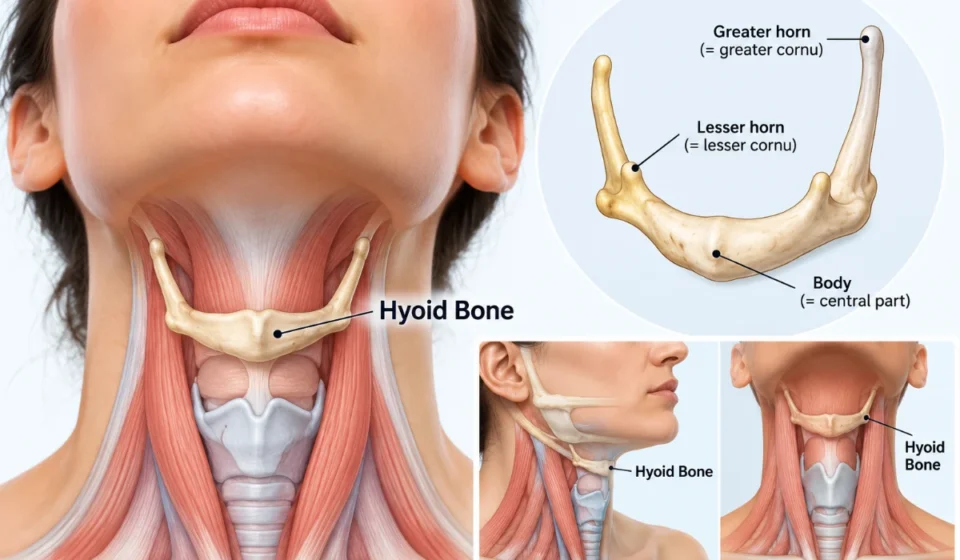

The hyoid bone is a small, U-shaped bone located in the front of the neck, between the lower jaw (mandible) and the thyroid cartilage of the larynx. Unlike most bones in the human skeleton, it does not directly articulate with any other bone. Instead, muscles and ligaments hold it in position, allowing it to move during swallowing, speaking, and tongue movements. It also contributes to the positioning and stability of the upper airway.
Although relatively small, the hyoid plays an important role in coordinating movements of the tongue, throat, and voice box. Its distinctive structure provides attachment points for several muscles involved in everyday activities. Understanding hyoid bone anatomy helps explain how these muscles work together to support swallowing, speech production, and normal movement of the larynx.
Where Is the Hyoid Bone Located?
The hyoid sits in the anterior neck, just below the mandible and above the thyroid cartilage. The thyroid cartilage forms the prominent part of the larynx commonly called the Adam’s apple. The bone lies near the base of the tongue and above the voice box, making it an important connection point between structures involved in speaking and swallowing.
In adults, the hyoid bone location is generally around the level of the fourth cervical vertebra (C4), although its position varies with age, posture, and movement. It can move upward, downward, and forward as surrounding muscles contract during activities such as swallowing.
Unlike the cervical vertebrae, which form part of the spinal column, the hyoid is suspended within the soft tissues of the neck. Its position allows the tongue and larynx to move together without requiring a rigid connection to another bone.
Read more: Hyoid Bone: Anatomy Location Functions and Pain Causes
Hyoid Bone Anatomy and Structure
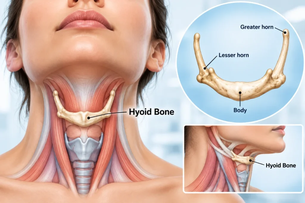
The hyoid bone structure consists of three main anatomical components: a central body, two greater horns, and two lesser horns. Together, these parts form a horseshoe-like shape that opens toward the back of the neck.
Each region provides attachment sites for muscles or ligaments involved in tongue movement, swallowing, and laryngeal function. The arrangement of these structures gives the bone both stability and flexibility.
Body of the Hyoid Bone
The body is the central, broad portion of the hyoid bone. It forms the front of the U-shaped structure and connects with the greater horns on either side.
Its anterior and posterior surfaces provide attachment areas for muscles associated with the tongue and larynx. Because it serves as a central attachment region, the body helps transfer muscular forces during swallowing and other movements.
Greater Horns (Greater Cornua)
The greater horns are two elongated projections extending backward and outward from the body. They form the longer arms of the U-shaped bone.
These projections provide attachment points for muscles and membranes associated with the tongue, pharynx, and larynx. Their arrangement helps distribute forces generated by surrounding muscles during coordinated movements.
Lesser Horns (Lesser Cornua)
The lesser horns are two smaller, cone-shaped projections situated near the junction of the body and greater horns.
They provide attachment points for the stylohyoid ligaments, which connect the hyoid region to the styloid processes of the temporal bones. These ligamentous connections help suspend the hyoid while allowing controlled movement.
| Anatomical Part | Structure | Main Significance |
| Body | Broad central portion | Provides important muscle attachment sites |
| Greater horns | Two long posterior projections | Support muscular and membranous attachments |
| Lesser horns | Two small projections | Provide attachment for the stylohyoid ligaments |
What Is the Function of the Hyoid Bone?
The primary hyoid bone function is to provide movable support for the tongue and larynx. Its muscular connections allow several structures in the neck to move together during swallowing, speech, and breathing.
Role in Swallowing and Tongue Movement
During swallowing, muscles above the hyoid contract, moving the bone upward and forward. This movement helps elevate the larynx and contributes to opening the upper esophageal sphincter, allowing swallowed material to pass toward the esophagus.
The movement also supports airway protection by helping coordinate laryngeal elevation with other swallowing mechanisms. The hyoid does not perform swallowing independently; its action is part of a coordinated sequence involving the tongue, pharynx, and larynx.
The bone also provides an important anchoring point for muscles associated with tongue movement and swallowing. These attachments help the tongue change position during chewing, speaking, and moving food through the mouth.
Role in Speech and Airway Support
Speech depends on precise coordination between the tongue, larynx, respiratory muscles, and surrounding tissues. The hyoid bone provides a movable attachment region that helps these structures adjust their positions during vocalization.
Its connection with the tongue and larynx supports movements needed for articulation and changes in vocal tract shape. However, it does not generate sound itself; sound production primarily involves airflow and vibration of the vocal folds.
Through its muscular connections, the hyoid also contributes to upper airway stability, particularly by helping maintain the positioning of the tongue and related soft tissues.
Muscles Attached to the Hyoid Bone
Several muscles influence the position and movement of the hyoid. They are commonly organized into two groups: suprahyoid and infrahyoid muscles.
These muscle groups work together to elevate, lower, or stabilize the bone depending on the activity being performed.
Suprahyoid Muscles
The suprahyoid muscles lie above the hyoid and connect it functionally with the mandible, skull, and floor of the mouth.
The four paired muscles are:
- Digastric: Helps elevate the hyoid and can assist in lowering the mandible when the hyoid is stabilized.
- Stylohyoid: Elevates and draws the hyoid backward during swallowing.
- Mylohyoid: Forms much of the muscular floor of the mouth and helps elevate the hyoid and tongue.
- Geniohyoid: Pulls the hyoid forward and upward, contributing to swallowing movements.
Together, these muscles help raise the hyoid during swallowing while supporting coordinated tongue and jaw movements. Their actions vary depending on whether the jaw or hyoid is stabilized.
Infrahyoid Muscles
The infrahyoid muscles are located below the hyoid, primarily in the anterior neck. They help lower or stabilize the hyoid and control movements of the larynx.
The four paired muscles include the sternohyoid, omohyoid, thyrohyoid, and sternothyroid.
The sternohyoid and omohyoid help depress the hyoid. The thyrohyoid can lower the hyoid or elevate the larynx, depending on which structure is stabilized. The sternothyroid acts on the thyroid cartilage rather than attaching directly to the hyoid.
These coordinated actions allow the hyoid bone and larynx to return toward their resting positions after movements such as swallowing.
Why Is the Hyoid Bone Unique?
The hyoid is unusual because it has no direct bony articulation with the rest of the skeleton. Most bones connect to neighboring bones through joints, but the hyoid is suspended by muscular and ligamentous attachments.
This arrangement allows controlled mobility while maintaining support for the tongue and larynx. The stylohyoid ligaments help suspend it from the skull, while surrounding muscles actively adjust its position.
Another distinctive feature is that it serves as a shared attachment point for muscles above and below it. The suprahyoid and infrahyoid groups work together to control its position, helping coordinate the movements required for swallowing and speech.
Frequently Asked Questions
1. What is the hyoid bone?
The hyoid is a small, U-shaped bone in the front of the neck. It supports the tongue and larynx through muscular and ligamentous attachments, helping coordinate swallowing, speech, and tongue movements.
2. Where is the hyoid bone located?
It is located below the lower jaw and above the thyroid cartilage of the larynx. In adults, it generally lies around the level of the fourth cervical vertebra (C4), although its exact position varies.
3. Why is the hyoid bone called a floating bone?
It is sometimes called a floating bone because it does not directly articulate with another bone. However, it is not freely floating; muscles and ligaments suspend and stabilize it within the neck.
4. What are the three main parts of the hyoid bone?
The three main components are the central body, paired greater horns, and paired lesser horns. These structures provide attachment sites for muscles and ligaments involved in tongue and laryngeal movements.
5. What muscles attach to the hyoid bone?
The principal suprahyoid muscles include the digastric, stylohyoid, mylohyoid, and geniohyoid. The sternohyoid, omohyoid, and thyrohyoid are infrahyoid muscles that also attach to it.
6. How does the hyoid bone help with swallowing?
During swallowing, surrounding muscles move the hyoid upward and forward. This helps elevate the larynx, contributes to airway protection, and assists with opening the upper esophageal sphincter so food or liquid can pass toward the esophagus.
7. Does the hyoid bone help with speaking?
Yes. It provides muscular support for movements of the tongue and larynx, helping adjust their positions during speech. It does not produce sound directly but contributes to the coordinated movements needed for articulation and vocalization.